pathfinder kit Search Results


88
Bio-Rad pathfinder c trachomatis direct specimen system
Analysis of MIL-100(Fe) nanoparticle accumulation in chlamydial inclusions using TEM imaging. (A) An image of chlamydial inclusion in cytoplasm of RAW264.7 cells infected with C. <t>trachomatis</t> . (B) Photograph of phagosome with nanoMOFs. (C) Co-localization of MIL-100(Fe) nanoparticles with C. trachomatis EBs within inclusions in RAW264.7 cells. (D) EDX spectrum analysis showing two major peaks of iron at 6.5 and 7.1 keV from the area with nanoMOFs. The peaks are not detected from the area without nanoparticles. (A)-(D) Black and white arrowheads indicate chlamydial EBs and MIL-100(Fe) nanoparticles, respectively. White stars indicate chlamydial inclusions. Abbreviations: N, nucleus; C, cytosol; P, phagosome; E, extracellular space. Scale bar is 500 nm.
Pathfinder C Trachomatis Direct Specimen System, supplied by Bio-Rad, used in various techniques. Bioz Stars score: 88/100, based on 1 PubMed citations. ZERO BIAS - scores, article reviews, protocol conditions and more
https://www.bioz.com/product/pathfinder+kit/Pathfinder+Chlamydia+DFA+(Complete+Kit+with+Controls)/bio_rxiv__2023__04__04__535539-168-7-14
Average 88 stars, based on 1 article reviews
pathfinder c trachomatis direct specimen system - by Bioz Stars, 2026-10
88/100 stars
  Buy from Supplier

90
DiscoverX corporation pathfinder kit
Analysis of MIL-100(Fe) nanoparticle accumulation in chlamydial inclusions using TEM imaging. (A) An image of chlamydial inclusion in cytoplasm of RAW264.7 cells infected with C. <t>trachomatis</t> . (B) Photograph of phagosome with nanoMOFs. (C) Co-localization of MIL-100(Fe) nanoparticles with C. trachomatis EBs within inclusions in RAW264.7 cells. (D) EDX spectrum analysis showing two major peaks of iron at 6.5 and 7.1 keV from the area with nanoMOFs. The peaks are not detected from the area without nanoparticles. (A)-(D) Black and white arrowheads indicate chlamydial EBs and MIL-100(Fe) nanoparticles, respectively. White stars indicate chlamydial inclusions. Abbreviations: N, nucleus; C, cytosol; P, phagosome; E, extracellular space. Scale bar is 500 nm.
Pathfinder Kit, supplied by DiscoverX corporation, used in various techniques. Bioz Stars score: 90/100, based on 1 PubMed citations. ZERO BIAS - scores, article reviews, protocol conditions and more
https://www.bioz.com/product/pathfinder+kit/pathfinder+kit/pm37238699-254-18-17
Average 90 stars, based on 1 article reviews
pathfinder kit - by Bioz Stars, 2026-10
90/100 stars
  Buy from Supplier

90
DiscoverX corporation pathfinder assay kit
Analysis of MIL-100(Fe) nanoparticle accumulation in chlamydial inclusions using TEM imaging. (A) An image of chlamydial inclusion in cytoplasm of RAW264.7 cells infected with C. <t>trachomatis</t> . (B) Photograph of phagosome with nanoMOFs. (C) Co-localization of MIL-100(Fe) nanoparticles with C. trachomatis EBs within inclusions in RAW264.7 cells. (D) EDX spectrum analysis showing two major peaks of iron at 6.5 and 7.1 keV from the area with nanoMOFs. The peaks are not detected from the area without nanoparticles. (A)-(D) Black and white arrowheads indicate chlamydial EBs and MIL-100(Fe) nanoparticles, respectively. White stars indicate chlamydial inclusions. Abbreviations: N, nucleus; C, cytosol; P, phagosome; E, extracellular space. Scale bar is 500 nm.
Pathfinder Assay Kit, supplied by DiscoverX corporation, used in various techniques. Bioz Stars score: 90/100, based on 1 PubMed citations. ZERO BIAS - scores, article reviews, protocol conditions and more
https://www.bioz.com/product/pathfinder+kit/pathfinder+assay+kit/pm23516330-65-10-13
Average 90 stars, based on 1 article reviews
pathfinder assay kit - by Bioz Stars, 2026-10
90/100 stars
  Buy from Supplier

Image Search Results


Analysis of MIL-100(Fe) nanoparticle accumulation in chlamydial inclusions using TEM imaging. (A) An image of chlamydial inclusion in cytoplasm of RAW264.7 cells infected with C. trachomatis . (B) Photograph of phagosome with nanoMOFs. (C) Co-localization of MIL-100(Fe) nanoparticles with C. trachomatis EBs within inclusions in RAW264.7 cells. (D) EDX spectrum analysis showing two major peaks of iron at 6.5 and 7.1 keV from the area with nanoMOFs. The peaks are not detected from the area without nanoparticles. (A)-(D) Black and white arrowheads indicate chlamydial EBs and MIL-100(Fe) nanoparticles, respectively. White stars indicate chlamydial inclusions. Abbreviations: N, nucleus; C, cytosol; P, phagosome; E, extracellular space. Scale bar is 500 nm.

Journal: bioRxiv

Article Title: NanoMOFs with Encapsulated Photosensitizer: Accumulation in Chlamydia trachomatis Inclusions and Antimicrobial Effects

doi: 10.1101/2023.04.04.535539

Figure Lengend Snippet: Analysis of MIL-100(Fe) nanoparticle accumulation in chlamydial inclusions using TEM imaging. (A) An image of chlamydial inclusion in cytoplasm of RAW264.7 cells infected with C. trachomatis . (B) Photograph of phagosome with nanoMOFs. (C) Co-localization of MIL-100(Fe) nanoparticles with C. trachomatis EBs within inclusions in RAW264.7 cells. (D) EDX spectrum analysis showing two major peaks of iron at 6.5 and 7.1 keV from the area with nanoMOFs. The peaks are not detected from the area without nanoparticles. (A)-(D) Black and white arrowheads indicate chlamydial EBs and MIL-100(Fe) nanoparticles, respectively. White stars indicate chlamydial inclusions. Abbreviations: N, nucleus; C, cytosol; P, phagosome; E, extracellular space. Scale bar is 500 nm.

Article Snippet: Chlamydial inclusions were visualized by staining with Pathfinder C. trachomatis Direct Specimen System (#30704, BIO-Rad) according to manufacturer’s recommendations.

Techniques: Imaging, Infection

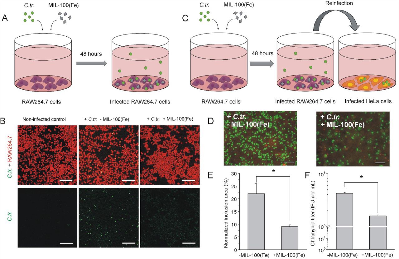
Intrinsic antibacterial effect of nanoMOFs. (A) The scheme of RAW264.7 cell infection and simultaneous treatment with MIL-100(Fe) nanoparticles. (B) After 48 hours of incubation, RAW264.7 macrophages were stained with Evans blue to visualize the cells (red) and FITC-labeled antibodies against chlamydial major outer membrane protein to visualize chlamydial inclusions (green). (C) The scheme of the experiment with reinfection of HeLa cells to determine infectivity of chlamydial progeny recovered from RAW264.7 macrophages, infected with C. trachomatis and treated or non-treated with nanoMOFs. (D) Images of reinfected HeLa cells (red) with chlamydial inclusions (green) visualized by staining with FITC-labeled antibodies against chlamydial major outer membrane protein. Scale bar is 50 µm for (B) and (D). (E) Quantitative analysis of reinfection efficacy determined as normalized chlamydial inclusion area. (F) Quantitative analysis of reinfection efficacy determined as infectious progeny yield. Data are shown as means ± SD. * p < 0.05, Mann-Whitney U test.

Journal: bioRxiv

Article Title: NanoMOFs with Encapsulated Photosensitizer: Accumulation in Chlamydia trachomatis Inclusions and Antimicrobial Effects

doi: 10.1101/2023.04.04.535539

Figure Lengend Snippet: Intrinsic antibacterial effect of nanoMOFs. (A) The scheme of RAW264.7 cell infection and simultaneous treatment with MIL-100(Fe) nanoparticles. (B) After 48 hours of incubation, RAW264.7 macrophages were stained with Evans blue to visualize the cells (red) and FITC-labeled antibodies against chlamydial major outer membrane protein to visualize chlamydial inclusions (green). (C) The scheme of the experiment with reinfection of HeLa cells to determine infectivity of chlamydial progeny recovered from RAW264.7 macrophages, infected with C. trachomatis and treated or non-treated with nanoMOFs. (D) Images of reinfected HeLa cells (red) with chlamydial inclusions (green) visualized by staining with FITC-labeled antibodies against chlamydial major outer membrane protein. Scale bar is 50 µm for (B) and (D). (E) Quantitative analysis of reinfection efficacy determined as normalized chlamydial inclusion area. (F) Quantitative analysis of reinfection efficacy determined as infectious progeny yield. Data are shown as means ± SD. * p < 0.05, Mann-Whitney U test.

Article Snippet: Chlamydial inclusions were visualized by staining with Pathfinder C. trachomatis Direct Specimen System (#30704, BIO-Rad) according to manufacturer’s recommendations.

Techniques: Infection, Incubation, Staining, Labeling, Membrane, MANN-WHITNEY

Photodynamic effect of MB-loaded nanoMOFs in C. trachomatis -infected RAW264.7 macrophages. (A) The scheme of the reinfection experiment with HeLa cells to determine an infectious progeny yield of infected RAW264.7 macrophages treated with different formulations, followed by light irradiation. (B) Images of reinfected HeLa cells (red) with chlamydial inclusions (green) visualized by staining with FITC-labeled antibodies against chlamydial lipooligosaccharide. Scale bar is 50 µm. (C) Quantitative analysis of the reinfection efficacy. To quantify an infection rate in HeLa cells, we normalized the area of inclusions to the area of HeLa cells. (D) Quantitative analysis of the reinfection efficacy determined as an infectious progeny yield. Data are shown as means ± SD. * p < 0.05, Mann-Whitney U test.

Journal: bioRxiv

Article Title: NanoMOFs with Encapsulated Photosensitizer: Accumulation in Chlamydia trachomatis Inclusions and Antimicrobial Effects

doi: 10.1101/2023.04.04.535539

Figure Lengend Snippet: Photodynamic effect of MB-loaded nanoMOFs in C. trachomatis -infected RAW264.7 macrophages. (A) The scheme of the reinfection experiment with HeLa cells to determine an infectious progeny yield of infected RAW264.7 macrophages treated with different formulations, followed by light irradiation. (B) Images of reinfected HeLa cells (red) with chlamydial inclusions (green) visualized by staining with FITC-labeled antibodies against chlamydial lipooligosaccharide. Scale bar is 50 µm. (C) Quantitative analysis of the reinfection efficacy. To quantify an infection rate in HeLa cells, we normalized the area of inclusions to the area of HeLa cells. (D) Quantitative analysis of the reinfection efficacy determined as an infectious progeny yield. Data are shown as means ± SD. * p < 0.05, Mann-Whitney U test.

Article Snippet: Chlamydial inclusions were visualized by staining with Pathfinder C. trachomatis Direct Specimen System (#30704, BIO-Rad) according to manufacturer’s recommendations.

Techniques: Infection, Irradiation, Staining, Labeling, MANN-WHITNEY